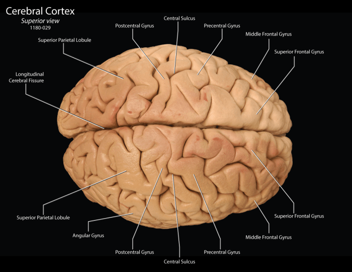
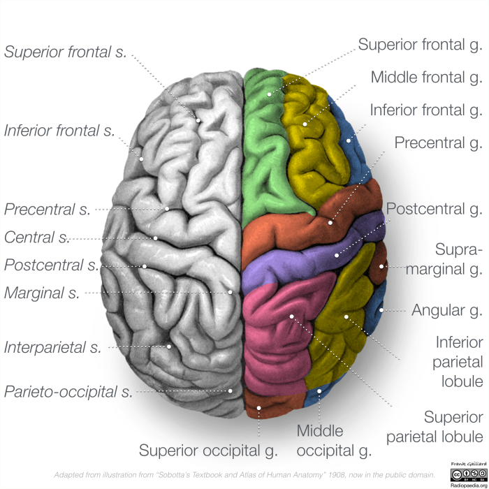
The superior view of the brain labeled unveils a complex and fascinating landscape, revealing the intricate architecture of the human mind. This perspective offers invaluable insights into the brain’s anatomical structures, blood supply, clinical implications, and imaging techniques, providing a comprehensive understanding of this vital organ.
From the major lobes to the sulci and gyri, the superior view dissects the brain’s topography, highlighting its functional regions. The arterial supply and venous drainage are meticulously mapped, showcasing the intricate network that sustains the brain’s metabolic demands.
Superior View of the Brain: Anatomical Structures

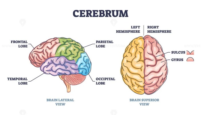
The superior view of the brain reveals several prominent anatomical structures. The brain is divided into two hemispheres, the left and right, separated by the falx cerebri. Each hemisphere is further divided into four lobes: the frontal lobe, parietal lobe, temporal lobe, and occipital lobe.
These lobes are separated by sulci, which are grooves in the brain’s surface.
Central Sulcus and Lateral Sulcus
Two prominent sulci on the superior view of the brain are the central sulcus and lateral sulcus. The central sulcus separates the frontal lobe from the parietal lobe and extends from the vertex to the lateral sulcus. The lateral sulcus separates the frontal and parietal lobes from the temporal lobe and runs parallel to the central sulcus.
Precentral Gyrus and Postcentral Gyrus
The precentral gyrus and postcentral gyrus are two gyri (ridges) located on the superior view of the brain. The precentral gyrus is located anterior to the central sulcus and is responsible for voluntary motor control. The postcentral gyrus is located posterior to the central sulcus and is responsible for sensory perception.
Blood Supply to the Superior Aspect of the Brain
The superior aspect of the brain is supplied by branches of the internal carotid artery and vertebral artery. The internal carotid artery gives rise to the anterior cerebral artery and middle cerebral artery, while the vertebral artery gives rise to the posterior cerebral artery.
Major Arteries and Their Branches
- Anterior cerebral artery:Supplies the medial surface of the frontal and parietal lobes.
- Middle cerebral artery:Supplies the lateral surface of the frontal, parietal, and temporal lobes.
- Posterior cerebral artery:Supplies the occipital lobe and medial temporal lobe.
Venous Drainage
The superior aspect of the brain is drained by the superior sagittal sinus, which runs along the midline of the brain, and the transverse sinuses, which run laterally from the superior sagittal sinus.
Clinical Significance of the Superior View of the Brain: Superior View Of The Brain Labeled

Lesions involving the superior aspect of the brain can have significant clinical implications. Damage to the precentral gyrus can result in paralysis or weakness on the contralateral side of the body, while damage to the postcentral gyrus can result in sensory loss on the contralateral side of the body.
Localizing Brain Tumors and Other Pathologies
The superior view of the brain can be used to localize brain tumors and other pathologies. For example, a tumor in the frontal lobe may cause personality changes or difficulty with executive function, while a tumor in the parietal lobe may cause difficulty with spatial orientation or attention.
Neurosurgical Procedures, Superior view of the brain labeled
The superior view of the brain is also important for neurosurgical procedures. Surgeons may need to access the superior aspect of the brain to remove a tumor, repair a blood vessel, or treat other conditions.
Imaging Techniques for Visualizing the Superior View of the Brain

Several imaging techniques can be used to visualize the superior view of the brain. These techniques include:
- Computed tomography (CT):Uses X-rays to create cross-sectional images of the brain.
- Magnetic resonance imaging (MRI):Uses magnetic fields and radio waves to create detailed images of the brain.
- Magnetic resonance angiography (MRA):Uses MRI to visualize blood vessels in the brain.
- Positron emission tomography (PET):Uses radioactive tracers to measure metabolic activity in the brain.
Comparison and Advantages
Each imaging technique has its own advantages and disadvantages. CT is a quick and inexpensive technique, but it provides less detailed images than MRI. MRI provides more detailed images, but it is more expensive and time-consuming than CT. MRA can be used to visualize blood vessels in the brain, which can be helpful for diagnosing and treating vascular disorders.
PET can be used to measure metabolic activity in the brain, which can be helpful for diagnosing and treating neurological disorders.
Question & Answer Hub
What are the major lobes visible from the superior view of the brain?
The frontal, parietal, occipital, and temporal lobes are all visible from the superior view.
What is the significance of the central sulcus?
The central sulcus separates the frontal and parietal lobes and serves as a landmark for identifying the primary motor and sensory cortices.
How is the superior view of the brain used in neurosurgery?
The superior view provides a roadmap for neurosurgeons to access deep-seated brain structures and remove tumors or other lesions.